Le site des ophtalmologistes de France
Encyclopédie de la vue
Vous êtes ici
Renforcement scléral
Renforcement scléral
pour myopie forte
Scleral reinforcement

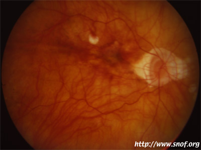
Myopie forte
Cliché Pr Mathis CHU Rangueil-Toulouse France
Généralités
Le renforcement scléral est une technique chirurgicale qui consiste à essayer de diminuer l'évolution d'une myopie et la fréquence des complications, en sanglant le pôle postérieur de l'oeil avec une bande de PTFE ou polytétrafluoréthylène (téflon). Cette technique s'adresse aux myopies fortes (au delà de -10 dioptries), avec staphylome.
Cette technique est peu utilisée en France.
Traduction du résumé de l'article de D. Chauvaud, de Paris:
"Le renforcement scléral est proposé pour stabiliser l'acuité visuelle de patients présentant un staphylome myopique maculaire. Bien que de nombreux patients aient été traités, cette procédure est encore controversée.
BUT Evaluation de la réalisation du renforcement scléral et du risque opératoire.
PATIENTS ET METHODE Les patients ayant bénéficié de cette technique prospective étaient ceux ayant présenté une baisse de la vision, un staphylome maculaire associé à des lésions atrophiques ou des ruptures de la bruch. Seize yeux de treize patients ont été opérés, avec une seule bande de PTFE.
RESULTATS Lors du dernier examen l'acuité visuelle fut inchangée pour 14 yeux. Dans un cas, une amélioration de la vision fut mise en rapport avec la disparition d'un décollement maculaire par raccourcissement de la longueur axiale. Dans un cas la vision a diminué, ce qui fut corrélé à un mauvais positionnement de la bande. Une diplopie apparut dans deux cas. Un décollement choroidien, avec une hémorragie du vitré disparut avec des séquelles.
CONCLUSION Une technique précise est nécessaire pour éviter le risque opératoire. Des études sur le long terme sont nécessaires pour évaluer le bénéfice du renforcement scléral."
Chauvaud D, Assouline M, Perrenoud F. [Posterior scleral reinforcement surgery] Service d'Ophtalmologie, Hotel-Dieu, Paris. J Fr Ophtalmol 1997;20(5):374-82.
Scleral reinforcement is proposed to stabilize the visual acuity in patients with macular myopic staphyloma. Although many patients have been treated, this procedure is still debated. PURPOSE: Evaluation of the feasibility of scleral reinforcement and the operative risk of this procedure. PATIENTS AND METHODS: Patients were eligible for this prospective study patients with a clinical history of visual loss, staphyloma concerning the macular area associated with atrophic lesion and or lacquer cracks. Sixteen eyes in 13 successive patients have been operated on with a single band of PTFE. RESULTS: At the last examination, visual acuity was unchanged for 14 cases. In one case, an improvement of the vision was related to the disappearing of a macular detachment by shortening of the axial length. In one case, vision decline was associated with inadequate band position. Diplopia occurred in 2 cases. A choroidal detachment, and a vitreous haemorrhage disappeared without sequelae. CONCLUSION: An accurate technique is necessary to avoid operative risk. Further long term studies are needed to assess the benefit of scleral reinforcement.
Bibliographie
Xu Y, Liu H, Niu T, et Al. [Long-term observation of curative effects of posterior scleral reinforcement surgery in patients with juvenile progressive myopia] Chung Hua Yen Ko Tsa Chih. 2000 Nov;36(6):455-8. Chinese.
Gerinec A, Slezakova G. [Posterior scleroplasty in children with severe myopia.] Bratisl Lek Listy. 2001;102(2):73-8.
Avetisov ES, Tarutta EP, Iomdina EN, Vinetskaya MI, Andreyeva LD. Nonsurgical and surgical methods of sclera reinforcement in progressive myopia. Acta Ophthalmol Scand. 1997 Dec;75(6):618-23.
Coroneo MT, Beaumont JT, Hollows FC. Scleral reinforcement in the treatment of pathologic myopia. Aust N Z J Ophthalmol. 1988 Nov;16(4):317-20.
Enculescu A. [Posterior scleral reinforcement in myopia--a harmless intervention?] Oftalmologia. 1991;35(3-4):85-6. Romanian.
Goldschmidt E. Myopia in humans: can progression be arrested? Ciba Found Symp. 1990;155:222-9; discussion 230-4. Review.
Jacob-LaBarre JT, Assouline M, Byrd T, McDonald M. Synthetic scleral reinforcement materials: I. Development and in vivo tissue biocompatibility response. J Biomed Mater Res. 1994 Jun;28(6):699-712.
Jacob-LaBarre JT, Assouline M, Conway MD, Thompson HW, McDonald MB. Effects of scleral reinforcement on the elongation of growing cat eyes. Arch Ophthalmol. 1993 Jul;111(7):979-86.
Karabatsas CH, Waldock A, Potts MJ. Cilioretinal artery occlusion following scleral reinforcement surgery. Acta Ophthalmol Scand. 1997 Jun;75(3):316-8.
Morelle N, Wery V, Croughs P. [Progressive myopia and posterior scleral reinforcement: retrospective studies] Bull Soc Belge Ophtalmol. 1996;262:43-5. French.
Riffenburgh RS. Dangers of scleral reinforcement for myopia. Ophthalmic Surg. 1985 Nov;16(11):730-1. No abstract available.
Thompson FB, Turner AF. Computed axial tomography on highly myopic eyes following scleral reinforcement surgery. Ophthalmic Surg. 1992 Apr;23(4):253-9.
Zhang J, Wu N. [Retinal detachment--severe complication after posterior scleral reinforcement operation] Chung Hua Yen Ko Tsa Chih. 1997 May;33(3):210-2. Chinese.


